Showing posts with label Hereditary Disease Foundation. Show all posts

Wednesday, October 23, 2024

At Harvard-MGH’s Mouro Pinto Lab, deploying CRISPR in the quest to cure Huntington’s disease


With a $1 million grant from the Hereditary Disease Foundation (HDF), a team of top researchers led by Ricardo Mouro Pinto, Ph.D., of Harvard Medical School, is deploying CRISPR to target genetic modifiers of Huntington’s disease.


In brain cells, these modifiers accelerate or slow so-called somatic expansions in HD.


The Mouro Pinto Lab at Harvard-affiliated Massachusetts General Hospital (MGH) specializes in research on these expansions. Scientists describe this process as the tendency of the mutant, expanded, disease-causing huntingtin gene to keep expanding abnormally. This causes the dysfunction – and possibly also death – of brain cells, leading to HD symptoms  (click here to read more). As scientists now see somatic expansion as a key driver of HD, research on this process has burgeoned.


Investigators have identified the modifier genes affecting expansion through deep research in human data. In speeding or slowing somatic expansion, those genes can hasten or delay disease onset. Many academic and biopharma groups, including the Mouro Pinto Lab, are investigating modifiers as potential drug targets.


The Mouro Pinto Lab is focusing on how one modifier gene, MLH3, acts harmfully as a “scissors” in HD. (By convention, human MLH3 gene is always capitalized and italicized; in contrast, the non-human gene is rendered as Mlh3.)


In an August 7 interview at his lab, Dr. Mouro Pinto described an experimental drug his team is testing in mice to deactivate the MLH3 scissors, a potential first step in seeking a treatment in people.


“I'm extremely optimistic about our approach,” Dr. Mouro Pinto said. “We can see what our drug does to somatic expansions. Can it slow it down? And our preliminary data is very encouraging in the sense that we can do that.”


On August 10, Dr. Mouro Pinto provided an update on the project at HDF’s HD2024: Milton Wexler Biennial Symposium. Announced in October 2023, the foundation’s two-year Transformative Research Award supports his team’s research of “therapeutic targeting of somatic CAG expansions with CRISPR base editing.” A group of anonymous donors funds the award.


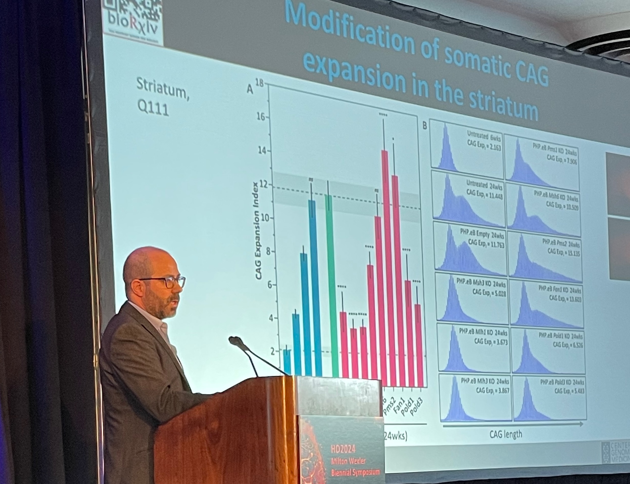
Above, at the HDF symposium, Dr. Mouro Pinto displays a slide demonstrating how his lab’s experimental drug slowed somatic expansion in the striatum of mouse brains. In humans, the 
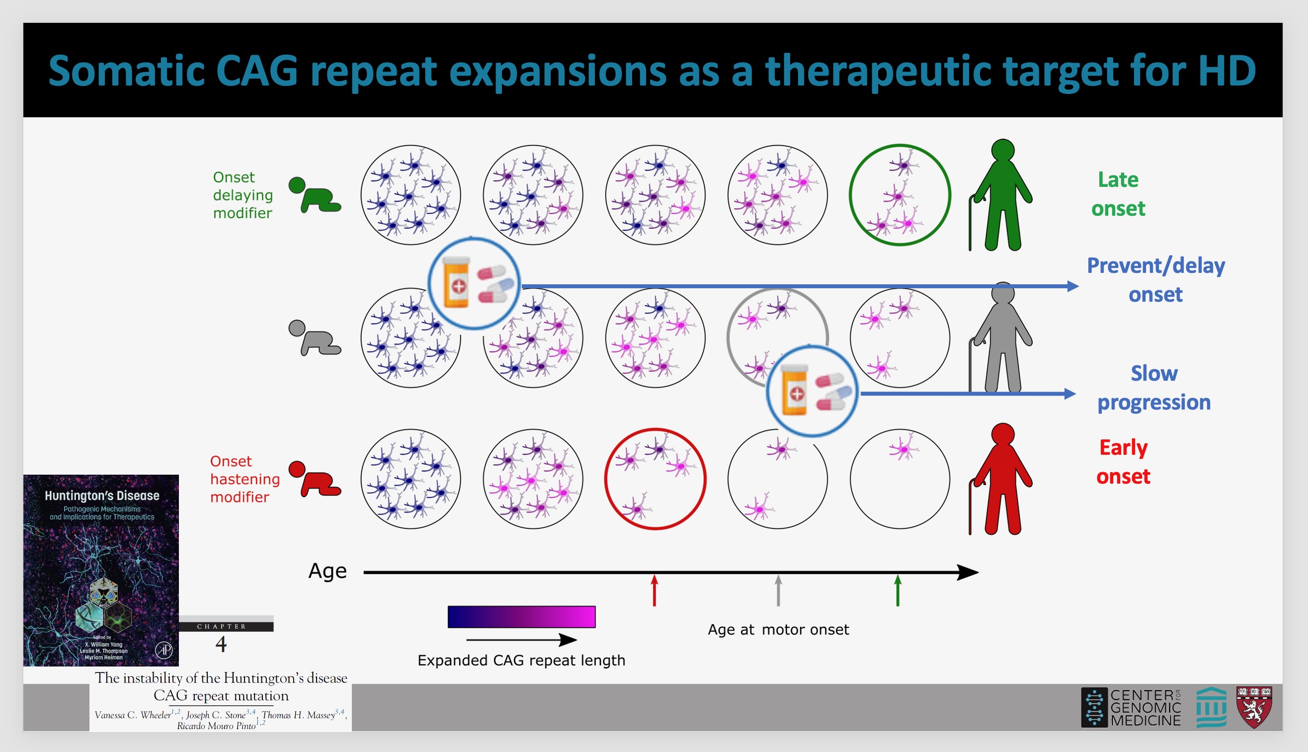
striatum is one of the areas most affected by HD. Below, a Mouro Pinto slide illustrates how a drug could impact somatic expansion and therefore prevent or delay HD onset, or slow progression of the disease in humans (photo of Dr. Mouro Pinto by Gene Veritas, aka Kenneth P. Serbin, image of slide courtesy of Dr. Mouro Pinto). (To make it larger, click on an image.)


Innovative collaborators


Seven innovative labs are collaborating with the Mouro Pinto team.


James Gusella, Ph.D., whose Harvard lab discovered the huntingtin gene in 1993, is one of three co-investigators on the project.


Gusella’s team has developed the human cell line that “allows us to model somatic instability so we can test the drugs,” Dr. Mouro Pinto explained.  That cell line has the expansions of the segment of the DNA code CAG (cytosine, adenine, and guanine), identified in 1993 as the underlying cause of HD.


At the symposium the HDF awarded Gusella the Leslie Gehry Prize for Innovation in Science, including $100,000 for research and a plaque with a small sculpture by the renowned architect Frank Gehry.


Specialists in somatic expansion


A native of Porto, Portugal, Dr. Mouro Pinto received his Ph.D. in molecular genetics at Brunel University in England in 2010, focusing on Friedreich’s ataxia, a debilitating genetic neuromuscular disorder. Along with HD, Friedreich’s is one of more than 50 repeat expansion disorders. In Friedreich’s and others, somatic expansion also plays a role. Scientists also refer to this process as somatic instability.


From 2010-2015, Dr. Mouro Pinto worked as a postdoctoral fellow in the lab of Vanessa Wheeler, Ph.D., an associate professor of neurology at Harvard, MGH researcher, and pioneer in the study of somatic instability. The Wheeler lab is collaborating with the Mouro Pinto group on the HDF project.


For Portuguese-speaking advocates, in 2022 I interviewed Dr. Mouro Pinto in his native tongue, discussing his work and outlook for HD therapies. We spoke at the 17th HD Therapeutics Conference, sponsored by CHDI Foundation, Inc., the largest private funder of HD research and a backer of the Mouro Pinto Lab.


On August 6, the HDF and MGH co-hosted a tour of the Mouro Pinto Lab for about 20 HDF officials, donors, and HD family members. Dr. Wheeler participated, too. I took part at the invitation of the HDF. At the start, Dr. Mouro Pinto presented an overview of the HDF project.


Above, before the tour of his lab, Dr. Mouro Pinto presents a slide demonstrating the path of his scientific studies. Below, Dr. Mouro Pinto explains the purpose of a PCR (polymerase chain reaction) workstation, where they isolate and make billions of copies of the CAG repeat so they can study it in more detail (photos by Gene Veritas). 


 A key task: measuring the expansion


In 2016 Dr. Mouro Pinto won the first Berman-Topper Family HD Career Development Fellowship from the Huntington’s Disease Society of America. That funding allowed him to conduct research demonstrating that modifier genes can speed or slow somatic expansion in mice, he said.


According to Dr. Mouro Pinto, his lab’s more recent examination of postmortem tissue from HD patients revealed that somatic expansion had occurred in about 30 different brain regions. In all, 50 different tissues were examined, showing different rates of somatic expansion in the liver, muscle, kidney, and others. Some of the fastest expansion was in the brain, he added.


To measure a drug’s effect in a clinical trial, brain biopsies are currently not an option, Dr. Mouro Pinto pointed out. However, measuring somatic expansion in blood, liver, and cerebrospinal fluid, which bathes the brain, could serve as biomarkers. A biomarker is a sign of a disease or effect of a drug. He noted that other researchers are already investigating somatic expansion in the blood.


“What is the right tissue, bio fluid to obtain from the patient?” Dr. Mouro Pinto asked. “And then what is the right test to be sensitive and accurate? So those two things are actually other aspects of research in our lab that we're trying to develop.”


Having those biomarkers will be crucial if the potential CRISPR drug reaches a human clinical trial, he observed.


CRISPR: a one-time, permanent edit of the gene


CRISPR stands for “clustered regularly interspaced short palindromic repeats,” a strand of RNA that, when activated by an enzyme, can edit DNA. Bacteria evolved this technique to defend against viruses.


Jennifer Doudna, Ph.D., and Emmanuelle Charpentier, Ph.D., won the 2020 Nobel Prize in Chemistry for their work in identifying and understanding CRISPR. Click here to read more about CRISPR, its potential for treating HD, and its powerful implications for the future of humanity.


Scientists have now used CRISPR to edit human genes in labs and in clinical trials that have resulted in drugs approved by the U.S. Food and Drug Administration (FDA). In December 2023 the FDA approved two CRISPR drugs for sickle cell disease, an inherited disorder that primarily affects people of African descent.


This first wave of CRISPR clinical trials has started with the “lowest-hanging fruit,” diseases that “affect the liver or the eye or the ear,” Dr. Mouro Pinto pointed out. The brain is far more difficult to research, so it has been challenging to address brain diseases with CRISPR drugs, he said.


“You're doing it at the level of the DNA and that causes a permanent change,” he emphasized. “Once you treat and you introduce the change you made, that will stay in that cell forever.”


A CRISPR clinical trial “will look, essentially, the same as any other HD trial,” he explained. “You'll need to collect samples. And you'll need to conduct a battery of physical tests, cognitive tests and behavioral tests. It will still be evaluated exactly by the same standards as any other clinical trial.”


In contrast with most other HD drug approaches, CRISPR has a key, beneficial difference. “It’s a one-time treatment,” Dr. Mouro Pinto said.


An ‘amazing toolbox’


Dr. Mouro Pinto underscored that in addition to serving as a drug, CRISPR has provided an “amazing toolbox” for less costly and more efficient and precise lab research.


In contrast with the old, very expensive process of breeding large numbers of mice over years, CRISPR “has accelerated the rate of research tremendously” and dramatically reduced the numbers needed, he said. With CRISPR, they can much more quickly pursue tasks such as making mutations in mice or screening a large number of genes to see which might modify somatic expansion, he said. The lab’s paper on this topic will be published in Nature Genetics.


The lab also can deliver a CRISPR reagent to a young mouse, transforming it into a model for study and producing results in a few weeks.


A unique ‘humanized Mlh3 mouse’


Use of CRISPR enabled a “really critical” step in the HDF project: creation of what Dr. Mouro Pinto described as a “humanized Mlh3 mouse,” a unique research step. To prepare the potential CRISPR drug for testing in mice for efficacy against HD and safety, the lab introduced into the animals a small sequence of human DNA from the MLH3 gene.


The scientists have crossed these mice with engineered HD mice. After a few generations downstream, this will result in mice with the expanded and the humanized modifier gene, which will be tested with the CRISPR reagent to see if it stops somatic expansion, Dr. Mouro Pinto continued.


The crossing was also necessary to assure that the humanized segment of DNA itself does indeed experience somatic instability, because the goal of the research is to stop instability, he added.


Another key step will involve measuring the CRISPR reagent’s impact on somatic expansion, irregular movements, and behavioral symptoms in the mice, Dr. Mouro Pinto said. For this stage, the lab will have the key assistance of the “extremely experienced” Cathleen Lutz, Ph.D., M.B.A., vice president of  The Jackson Laboratory Rare Disease Translational Center, he noted.


‘Promoting the flavor without the scissors’


As it develops greater precision, the Mouro Pinto Lab has found that one of two MLH3 variants has the scissors that cause harmful genetic cutting. That gets closer to solving the HD puzzle.


In a February HDF webinar about the project, Dr. Mouro Pinto explained that in people without HD, the MLH3 variant without scissors – the good variant – is present.


He said that he is confident that promoting expression (activation) of the good variant over the bad would be better tolerated as a treatment than simply turning off the bad. He described this approach as “promoting the flavor without the scissors, as opposed to completely getting rid of the protein” that results from the gene.


“In a mouse that doesn’t have the scissors, you completely stabilize the repeat,” Dr. Mouro Pinto stated in the webinar. “We know that the scissors component of this protein is essential for promoting CAG expansions.”


Furthermore, in human HD cells the team not only reduced but eliminated the bad version of MLH3.


They achieved this using a technique known as base editing.


Dr. Mouro Pinto noted that with standard CRISPR editing, a sequence of DNA can be broken up, potentially causing unwanted effects. In contrast, in base editing, no breakage occurs, because scientists edit the DNA by simply changing a letter of the genetic code, for example, from A (adenine) to G (guanine), or C (cytosine) to T (thymine).


Significantly, his team edited both copies of the MLH3 gene and completely shifted expression from the “bad” version towards only making the “good” “scissor-less” version of MLH3 protein. As a result, the experiment completely stabilized the CAG repeat (i.e. the CAG stopped expanding in edited cells), Dr. Mouro Pinto stressed.


There are two bases because, as Dr. Mouro Pinto reminded, every cell has two copies of each gene – a copy from each parent.


Partnering on more precise gene editing


Only a few projects have started exploring base editing for HD, and most are happening in research labs such as his, Dr. Mouro Pinto said.


To maximize the benefits for HD patients, the HDF project will seek to improve on this editing.


Recognizing the many other labs are examining directly targeting the CAG expansion, Dr. Mouro Pinto believes that, instead, deactivating a modifier gene such as the MLH3 scissors is a safer and easier strategy.


On this aspect, the Mouro Pinto Lab will partner with co-investigator David Liu, Ph.D., of both Harvard and the Massachusetts Institute of Technology (MIT). Dr. Liu invented both base editing and prime editing.


As Dr. Mouro Pinto pointed out in our interview, base editing allows for more “precise base changes.” In the HDF webinar, he noted that this method prevents breaking up of the DNA.


Harvard’s Benjamin Kleinstiver, Ph.D., another innovative co-investigator, has engineered CRISPR enzymes that “literally can go anywhere in the genome,” Dr. Mouro Pinto added. This enables the team to target any sequence of DNA it needs to, he said.


The lab continues to seek improvements in its enzyme to “achieve highest specificity and maximum efficacy,” he said.


The potential path to a clinical trial


“It's too early” to gauge whether the experimental CRISPR reagent can undergo testing in humans and have a “therapeutic impact,” Dr. Mouro Pinto told me, noting that four key questions must be answered in mice first. The lab aims to answer them in the coming months.


He described the outcome needed in the series of mouse experiments: “Did we change the DNA? Yes. Okay. Did we change which version of MLH3 is made? Yes. Okay. Next. Did we reduce the CAG instability? Yes. And then do we have any impact on HD symptoms?”


Currently the lab is working on the first step, with “promising” indications so far, Dr. Mouro Pinto said.


If the project proves successful, other, distinct projects testing the reagent in other animals, such as nonhuman primates, would follow, Dr. Mouro Pinto explained. The HDF grant does not include funds for those steps.


Emphasizing safety, avoiding unwanted edits


Dr. Mouro Pinto underscored that the team strives to find the safest CRISPR drug possible.


In line with more stringent FDA standards and bioethical concerns regarding gene editing, the project must carry out “due diligence” to avoid “very serious adversity.” That includes so-called off-target effects of gene editing, in which a gene such as one for cancer is accidentally activated or turned off, Dr. Mouro Pinto cautioned.


“It is not uncommon for these drugs to have some activity in unwanted regions of the genome,” he explained.  “We need to spend a lot of time looking for unwanted modifications.”


The CRISPR agent is “not yet ready for the clinic,” Dr. Mouro Pinto added.


He noted that the project is not doing edits in the sex cells; offspring therefore cannot inherit any genetic changes.


Finding the best way to deliver the cargo


The lab has no name yet for the experimental reagent, that is, its CRISPR enzyme.


“This is still an experimental reagent,” Dr. Mouro Pinto stressed. “I don't want to create false expectations. We are primarily putting effort into making sure that our cargo is good, that it's really doing what we want.”


The “cargo,” the potential drug, needs to be delivered safely and effectively into the brain and to the right cells, Dr. Mouro Pinto said. A common strategy in gene therapy is using a virus, specifically, an adeno-associated virus (AAV).


The Mouro Pinto Lab is using an AAV that works well in mice but not possible for humans, he said. As part of the HDF project, the team is searching for the ideal delivery system, which could be an AAV, a lipid nanoparticle, or extracellular vesicle, he continued. All three are tiny.


“There are many people now working on AAVs that you inject systemically,” Dr. Mouro Pinto said. “You give it into a vein, they go everywhere in your body including crossing the blood-brain barrier and entering the brain.” They can reach “almost every single neuron in the brain,” he added.


The blood-brain barrier is a membrane that protects the brain from harmful substances and germs.


Another project collaborator, Benjamin Deverman, Ph.D., of Vector Engineering and Harvard and MIT, has greatly improved the ability of AAVs to cross the blood-brain barrier in humans. In May, these critical findings for solving brain disorders were published in Science magazine.


“It's going to unlock this sort of roadblock that we have with delivery,” Dr. Mouro Pinto said of this breakthrough.


Lipid nanoparticles also can be injected into the blood. Some researchers are exploring oral administration of extracellular vesicles.


These vehicles pose less burden on clinical trial participants and patients in comparison with other methods, such as spinal taps or direct injection into the brain, a “complex surgical procedure,” Dr. Mouro Pinto observed.


“We're a little bit agnostic to the delivery strategy,” Dr. Mouro Pinto said, noting that the science of these delivery methods is evolving rapidly. By the time the project concludes in October 2025, “there might be a variety of different delivery options that we may want to consider.”


“Synergizing” with other HDF awardees


Dr. Mouro Pinto sees “opportunities to synergize” with the team that also received a 2023 Transformative Research Award, under the leadership of Beverly Davidson, Ph.D., of the University of Pennsylvania and Jang-Ho Cha, M.D., Ph.D., the chief scientific officer of Latus Biosciences. Latus focuses on precision delivery of gene therapy. An expert in AAVs, Dr. Davidson presented the team’s work at the HDF symposium.


Titled “Advancing gene therapies for HD” and focusing on AAVs, that project could potentially provide a delivery system for the CRISPR reagent, Dr. Mouro Pinto said.


Dr. Beverly Davidson presenting her team’s work on AAVs at the 2024 HDF symposium (photo by Gene Veritas).


Getting to market, looking beyond HD


In the event of the CRISPR reagent’s success in the lab, MGH will assist in commercializing it, Dr. Mouro Pinto said.


The hospital could license the technology to a biopharma company or, as in the case of the Davidson-Cha project, to start a company like Latus to bring the drug through a clinical trial and to market.


“We're open to those conversations and we've been fortunate to have a very collaborative interaction with industry partners so far,” Dr. Mouro Pinto told me.


The problem of somatic expansion “is shared across a large number of repeat expansion diseases,” he observed. “Individually, they're rare diseases. Collectively, they're not a rare disease. They actually affect a large number of patients around the world.”


“If our hypothesis is correct, the therapeutic benefit will not be limited to HD patients,” he concluded.


Disclosure: the Hereditary Disease Foundation covered my travel expenses to tour the Mouro Pinto Lab and attend the 2024 symposium.


Sadly, Michael McCabe, a 62-year-old Boston HD man who told his story at this year’s HDF symposium, died suddenly on September 12. Donations in Michael’s memory are suggested to the Huntington’s Disease Society of America.


Gene Veritas (left) and Dr. Mouro Pinto in the MGH lab (personal photo)

Thursday, August 15, 2024

At HDF symposium, a Huntington’s disease ‘hero’ who prays for scientists to find a cure


Recognizing the invaluable input from people living with Huntington’s disease, the Hereditary Disease Foundation (HDF) featured a conversation with Michael, a 62-year-old HD-affected Boston man, at its biennial conference of scientists seeking therapies for this incurable disorder.


Michael was interviewed about his HD symptoms by neurologist Diana Rosas, M.D., of Harvard University and Massachusetts General Hospital.


Titled “Living with Huntington’s Disease: Family Perspectives,” this HDF tradition of focusing on an HD-affected person took place on August 8 during HD2024: Milton Wexler Biennial Symposium. Convening some 300 researchers, biopharma officials, and advocates, the event ran August 7-10 at the Royal Sonesta Boston Hotel in Cambridge, MA.


HD usually impedes speech. I saw that affecting my mother. She died of the disorder at 68 in 2006, after two decades of symptoms, and I carry the HD gene.


Michael struggled but persistently formed words and sentences. “I pray for everybody,” Michael said, referring to the quest for therapies, during the Q&A after the interview.


Michael’s former wife attended in support of his advocacy, as did his two sons, both in their 20s.


Michael (left), who has Huntington's disease, and his physician, Diana Rosas, M.D. (photo by Gene Veritas, aka Kenneth P. Serbin)


A diagnosis in 2017


Born in Chicago, Michael grew up in Princeton, NJ. As a young adult he moved to Boston, where he studied to become a French chef. He spent a year traveling through France to master his profession. He worked in several restaurants in Boston and also at Gillette Stadium for the NFL’s New England Patriots.


Michael believes his father had HD, although he was never formally diagnosed, due to the limited knowledge about the disease as Michael grew up in the 1970s. His father was also an alcoholic. Michael’s aunt also suffered from HD and went into a care home.


Michael was diagnosed with HD in 2017.


It became ‘too dangerous and messy’ to cook


Dr. Rosas is Michael’s physician. As she noted, many lab researchers have little contact with HD-affected individuals. The interview aimed to inform them of the complex triad of symptoms and many psychosocial challenges posed by HD.


Dr. Rosas asked Michael to address questions about the first type of symptoms: movement disorders, including involuntary movements.


These symptoms, Michael explained, caused him to stop cooking: it had become “too dangerous and messy.” It also became harder to dress himself.


Typical of HD patients (including my mother), Michael has suffered several serious falls, leading to a broken wrist, ribs, neck, a punctured lung, and a subdural hematoma (a serious injury to the head). Though he had participated in research conducted by Dr. Rosas, the hematoma has prevented him from participating in clinical trials, because of a restriction by pharmaceutical companies.


“I like helping out however I can,” he said of his participation in research.


Michael, who lives alone, does have a chocolate labrador retriever that he walks.


Michael used to drink alcohol daily and smoke heavily. The drinking caused one of his falls, he said. He quit both habits. Alcohol was a “big part” of his life, he recalled, adding that he doesn’t “miss the days of drinking.”


A greatly modified daily routine


Dr. Rosas brought up another part of the HD triad: cognitive loss, executive dysfunction, and failing memory.


Michael observed that his loss of executive function prevented him from cooking, which had required preparing items and “lots of multitasking.”


Though he “can remember my bank card number,” he has ongoing difficulties with memory. He pays his cable and phone bills but has an accountant to assist with his overall finances. He still cares for two salt-water fish tanks, an activity he took up in his 20s.


Michael arises at 6 a.m., when he takes his medications: risperidone, an antipsychotic, twice daily; deluxotine for depression; and a multi-vitamin. He also takes medical marijuana.


After some small accidents, Michael stopped driving, now relying on Uber.


Overcoming impulsiveness and depression


Regarding the third part of the triad, psychiatric and mood disorders, Dr. Rosas observed that HD-affected individuals can become fixated or impulsive.


Michael agreed that this has affected him, recalling that his drinking also led him to be “very impulsive.” He also suffers from depression. Many HD-affected people become angry when faced with unexpected changes in their daily routine. Michael has also experienced this type of anger. Getting over the anger can take time, he added.


Like many of the affected, Michael also has difficulties sleeping. His drinking had exacerbated this problem.


“It’s like your mind and body are always on with HD,” he observed.


Indeed, HD-affected individuals burn lots of calories. Dr. Rosas recommends five meals per day, although Michael said he eats three to four. 


Dr. Rosas interviews Michael about his HD symptoms (photo by Gene Veritas).


‘You are a hero!”


In the Q&A following the interview, Michael expanded on aspects of his life.


One has involved his relationship with his ex-wife and sons. Michael said that the divorce occurred around the time of his diagnosis and was “probably” the result of it.


Michael saluted his former spouse as “one of my huge supporters. I haven’t had a girlfriend after my divorce. We were married for 24 years.”


He said that he has “two great kids” who are “successful and happy.”


Michael also socializes with friends, some of them also divorced.


Asked about the work of the researchers, Michael said, “I love them to death.” He added that he is looking forward to new advances.


Dr. Rosas asked what most worries Michael about HD.


“I suppose going to a home, going to an assisted living situation,” he said.


His capacity to manage on his own prompted praise. “You are a hero!” declared Tacie Fox, a family advocate and co-trustee of The Fox Family Foundation (which supports HD research), leading the audience to applaud enthusiastically.


“It feels like you have somehow navigated in a way that brings you joy in your life,” she added. “We’re struggling with that with my little sister. She watches a lot of TV. I’m in awe that you, living on your own, have marshaled that inner strength.”


The key role of modifier genes


At 64, I have been extremely fortunate to have not been diagnosed with HD. It is likely that I have benefited from modifier genes and other factors.


Like the rest of the audience, I was deeply moved by Michael’s courage and perseverance in living with HD.


I hope that when the inevitable symptoms arrive, I will have the same strength as Michael.


Stay tuned for upcoming articles on the conference proceedings, including deep discussion of the key role of modifier genes in the search for therapies.


Disclosure: the Hereditary Disease Foundation covered my travel expenses.

Monday, May 13, 2024

Aiming for multiple targets for Huntington’s disease therapies: a hopeful report from the Yang lab at UCLA


This article is in commemoration of Huntington’s Disease Awareness Month (May).


One of the most impactful university labs focusing on Huntington’s disease, the X. William Yang Research Group at the University of California, Los Angeles (UCLA) employs a multi-pronged approach to investigating potential therapies for this deadly brain disorder.


Started in 2002 by X. William Yang, M.D., Ph.D., the lab has produced several key findings on HD, mainly through the study of genetically modified (i.e., transgenic) mice, engineered to carry the HD mutation and exhibit some of the disease-like phenotypes (characteristics).


Dr. Yang was inspired to focus on Huntington's disease because of his interaction with patients in Venezuela – the world’s largest clusters of HD families – and the HD scientists working there. In 2000 and 2002 he was invited to observe these families and assist with studies by Nancy Wexler, Ph.D., the president of the HD-centered Hereditary Disease Foundation (HDF) and leader of the landmark effort to identify the HD gene in 1993.


Dr. Yang's Venezuela experience cemented his resolve to study HD in his own lab. Indeed, the first research grant ever received by Dr. Yang was from HDF. Today he serves as its scientific advisory board’s vice chair.


Dr. Yang’s team has also collaborated with CHDI Foundation, Inc., the largest private funder of HD therapeutic research. Pharmaceutical firms such as Roche (the world’s largest) and Ionis Pharmaceuticals, Inc., the developer of the Roche drug now in its second HD clinical trial, have consulted Dr. Yang for his expertise.


Dr. Yang has emerged as a leading academic voice in HD science. Listed as the first author, in February he and two other important prominent HD researchers – Leslie Thompson, PhD., of UC Irvine and Myriam Heiman, Ph.D., of the Massachusetts Institute of Technology (MIT) – published a major co-edited book. Huntington’s Disease: Pathogenic Mechanisms and Implications for Therapeutics presents the latest work on the disease’s medical impact, genetics, the huntingtin protein, new tools and models for research, and an overview of therapeutic approaches and clinical trial programs.


The back and front covers of Huntington’s Disease: Pathogenic Mechanisms and Implications for Therapeutics (image courtesy of Dr. Yang) (Click on an image to enlarge it.)


‘The stars are aligned’ for developing HD treatments


Although the use of human data in HD research has increased dramatically, crucial research in mice has become more relevant to potential therapies because of new biotechnologies and the availability of so-called “big data” made possible by powerful computing systems.


“This is completely unprecedented in terms of the kind of study we can do,” Dr. Yang told me in a 40-minute interview on January 29, noting the advantages of a “21st century toolbox.” “Mouse models in this context are extremely useful.”


We met in Dr. Yang’s office in his lab, which is located in UCLA’s Gonda (Goldschmied) Neuroscience and Genetics Research Center. I was invited to Los Angeles to offer my perspective as an HD gene carrier on the first day of a two-day HDF scientific workshop, co-chaired by Dr. Yang.


“I know it's probably an oxymoron to say that it's time to be hopeful, because we’ve been to a hopeful stage many times before,” Dr. Yang said, acknowledging the negative results of some recent clinical trials. He added that “the stars seem to be aligned” for developing HD treatments.


Dr. Yang (left) with project scientist Chris Park, Ph.D. At the far left is a confocal microscope, which uses laser light to obtain high-resolution images of thick tissues. Behind the men is a light sheet microscope, also used for obtaining high-quality images of tissues (photo by Gene Veritas, aka Kenneth P. Serbin).


Focusing on the brain


Dr. Yang grew up in Tianjin, China, a port city located 80 miles from the capital, Beijing. In 1985, Dr. Yang was one of five students selected by the Chinese government to participate in the Rickover Science Institute, founded by Admiral Hyman G. Rickover to foster high-school science education for both domestic and international students. Rickover developed the first nuclear-powered engines and first atomic-powered submarine.


“I did a whole summer of research at the NIH [National Institutes of Health], working on signaling pathways in rat brains,” Dr. Yang wrote in a follow-up e-mail to our interview. “The research experience got me really interested in studying the mammalian brain.”


The Rickover program is now called the Research Science Institute (RSI). Among other prestigious alumni are Harvard University’s Steve McCarroll, Ph.D., a leading molecular geneticist who also works on HD; and MIT/Broad Institute's Feng Zhang, Ph.D., a CRISPR research pioneer.


After RSI, Dr. Yang briefly studied at Peking University, one of China's top universities, before transferring to Yale University, where in 1991 he completed the highly demanding joint B.S./M.S. program in molecular biophysics and biochemistry.


Over lunch Dr. Yang and I reminisced about our years at Yale. I was privileged to graduate from Yale in 1982. I told Dr. Yang that I had seen Admiral Rickover give a public lecture at the university – a poignant moment for me as a history major because of his military and scientific prominence. I told Dr. Yang of my interest in tracking the contributions of Yale and its graduates like him to HD science and medicine (click here, here, and here to read more.)


Dr. Yang completed the joint M.D./Ph.D. program at The Rockefeller University (Ph.D., 1998) and Weill Medical College of Cornell University (M.D., 2000) in New York City. In 2002, he finished postdoctoral research in the Rockefeller lab of Nathanael Heintz, Ph.D., which focuses on HD and other neurological and psychiatric disorders.


Gene Veritas (left) (aka Kenneth P. Serbin) with Dr. William Yang in his UCLA office. In the background: a mouse medium spiny neuron. In humans this neuron is one of the cells most affected by Huntington’s disease (photo by Nan Wang, Ph.D., of the Yang Research Group).


A ‘trustworthy and versatile’ invention


Dr. Yang and his lab have made key contributions to HD science, including understanding the causes and potential pathways to therapies. The team also studies Alzheimer’s disease, Parkinson’s disease, and other neurodegenerative disorders.


As a Ph.D. student, Dr. Yang co-invented with Dr. Heintz and Peter Model, Ph.D., the first method to engineer Bacterial Artificial Chromosomes (BACs) to generate transgenic mice. BACs have the advantage of holding long strands of DNA with key regulatory elements that confer accurate gene expression in transgenic animals.


In an analysis of this research, which Drs. Yang, Model, and Heintz published in 1997, one leading biologist described their technique as “trustworthy and versatile” for cloning genes and the key task of learning the specific function of particular genes.


Indeed, scientists have used this method to generate a variety of transgenic animals, from zebrafish to mammals (click here to read more).


The key BACHD mouse


In 2008, Dr. Yang and other researchers published the results of a project creating the first BAC transgenic mouse model of HD, the BACHD mouse, their term for this mouse specifically engineered to study HD.


As Dr. Yang explained in our interview, the team inserted a long strand of a mutant (irregularly expanded) human huntingtin gene into the mice. Those genetic characteristics do not normally exist in mice. As they hoped, the mice developed dysfunction, displaying impaired movements, shrinkage to the same brain regions affected in HD, and damage to the synapses (the connections between brain cells).


“We developed different versions of these mouse models to allow us to ask, for example, which cell types in the brain with mutant huntingtin are important,” Dr. Yang said.


The team demonstrated the presence of mutant huntingtin in two key areas of the brain: medium spiny neurons in the striatum and pyramidal neurons (brain cells) in the cortex. (See the photo above with Dr. Yang, me, and an image of a medium spiny neuron. Also see the photo in the next section.)


In mice, humans, and other mammals, the cortex handles important processes such as cognition, memory, motor control, and sensory processing. The striatum – an area deep in the brain and greatly affected in HD – controls motor (movement), motor and reward learning, and executive function. In humans, this region is also known as the caudate and putamen. The Yang lab also examines communication between these regions.


Dr. Yang (left) and Nan Wang, Ph.D., a project scientist focusing on Huntington’s, in the lab (photo by Gene Veritas)


Using mice and genetics to understand HD


In detecting the impact of mutant huntingtin in those areas, that initial BACHD research revealed disease phenotypes in both striatum and cortex, Dr. Yang recalled. “That study turned out to be really important because, for the longest time, people thought the striatum, the medium spiny neuron, was really the primary site of action.”


Removing the mutant huntingtin from the cortex led to improvement in the mice’s behavior and even partially helped the striatum, Dr. Yang explained. Likewise, deleting mutant huntingtin from the striatum brought some improvement.


“But most importantly, if you reduce mutant huntingtin in both cortex and striatum, the BACHD model looks really, really good, almost as good as a normal mouse,” he added.


The BACHD work, he recalled, helped to convince the field that  the cortex is one of the key brain regions that should be targeted in HD clinical trials. The Roche/Ionis ASO lowers the level of huntingtin protein more in the cortex than in the caudate/putamen, according to preclinical studies in non-human primates.


In sum, Dr. Yang said, the studies of BACHD mice represent a “proof of concept that we can use this kind of a sophisticated – genetically as accurate as we could get – type of mouse model to inform about disease pathogenesis” – how HD develops, progresses, and, significantly, might be treated.


A mouse medium spiny neuron (image courtesy of Dr. Yang)


From disease switch to vulnerable neurons


The Yang Research Group has achieved other key findings, some in collaboration with other labs.


The Yang lab teamed with researchers at UC Irvine, UC San Francisco, the University of Pittsburgh, and the University of Tennessee to study the chemical modification of the huntingtin protein itself. This research focused on so-called “chemical tags” that naturally attach to the very beginning of the huntingtin protein, a small region acts like a disease switch.


In one experiment, this research used a BACHD-like mouse to mimic the tagging. That resulted in mice that had “very little disease despite having the HD mutation,” Dr. Yang explained. The results were published in 2009.


In 2015, the Yang Research Group published a separate study showing the genetic switch is necessary to prevent severe disease including neuronal loss and movement deficits, phenotypes reminiscent of those found in HD. These studies showed that the huntingtin protein itself and its chemical tags could be a source of new targets to develop therapies, Dr. Yang said.


From watching mice in ‘log rolling contests’ to unbiased genetic analysis


In 2013, the Federal Government announced the launch of the BRAIN initiative to enhance understanding of the human brain. The Yang lab was one of the first 59 in the country to receive support in the initial round of BRAIN funding. It now has its third grant. It receives support from other government agencies, as well as the HDF and CHDI. The lab’s achievements include developing a new, genetic way to label the complete, intricate shape of single brain cells, which allows the study of their function and dysfunction in diseases such as HD.


With big data and "the 21st century toolbox," the field of HD research has advanced from more traditional ways of observing diseased mice to more nuanced molecular, cellular and systems biology analyses, Dr. Yang explained.


In earlier research, by primarily relying on the behavior and pathology of individual mice, the work resulted in “relatively few readouts” of data, Dr. Yang observed. With that methodology, scientists had mice doing activities such as “spontaneously move” in an open area or on a rotarod, “like the ESPN log rolling contest,” he said. Scientists also routinely measured loss of brain matter.


Now, scientists can do a “big-scale, unbiased molecular studies” by examining tens thousands of datapoints, including analysis of DNA, RNA and proteins, Dr. Yang added.


Clues from gene expression about neuronal vulnerability


Collaborating with CHDI, the Yang lab’s work in this area has involved study of HD’s impact in different areas of the brain, moving beyond the standard understanding that most damage comes in the striatum. The lab has done this research using different types of engineered HD mouse models carrying different lengths of CAG repeats and measured the levels of tens of thousands of RNA transcripts ("RNA-seq," that is, RNA sequencing) in the mouse brains and peripheral tissues.


Published in 2016, the results noted that despite the presence of the mutant HD gene throughout the body, the disruption in gene expression in these HD mice is highly selective to the striatum, the most affected brain region in HD. The severity of the disruption is correlated with the length of CAG repeats in these mice. Moreover, the molecular defects in the striatum appear in young adulthood, worsening with age.


“There's about 100 or so genes that have essential function selective to the striatal neurons that are most affected in Huntington’s disease,” Dr. Yang said. “And somehow the mutant huntingtin knows to go there and make them the sickest, which we thought was a remarkable find – a sense that there's some fundamental mechanism connecting this CAG expansion to selective neuronal vulnerability.”


‘Perturbing’ the mice to understand human modifier genes


Taking advantage of the gene signatures from RNA-seq studies, especially those selectively disrupted in the striatum, the Yang lab embarked on a study using such gene signatures to sensitively detect "modifiers" of the disease. To achieve this, they used these genes to genetically “perturb” the mice, Dr. Yang explained.


“We basically genetically perturb the huntingtin mouse and say, ‘which gene, if we perturb them just right, can make the disease worse – that's one thing that's interesting – but more importantly make them better. And if better, how much better.’”


Continuing this line of work, the lab has continued testing the impact of other genes. These experiments include study of some of the human HD modifier genes – about ten – previously identified by the Genome Wide Association Study (GWAS) from over 9,000 HD-affected individuals and their relatives. The modifiers found by the Genetic Modifiers of Huntington's Disease Consortium can delay or hasten HD onset.


In addition, the Yang lab tested over 100 other candidate modifier genes identified in the prior systems biology work.


The scientists have tested large number of genetic mutants in HD mice to determine whether this makes the disease better or worse, Dr. Yang said. Noting that the results are still unpublished, Dr. Yang said that the team is drilling down on discovering the best gene targets that could help advance therapies to alleviate the disease.


Three potential ways to treat HD


Dr. Yang also discussed his outlook for therapies to slow, prevent, or reverse the course of Huntington’s. As noted, he believes that “the stars seem to be aligned” for the development of treatments.


In exchanging ideas with other HD scientists, he proposed the model of a stool – which needs four legs to remain stable –  as a metaphor for the benefit of developing multiple therapies (polypharmacy) that could act synergistically for HD.  


“If one drug could work for HD, that will be great. However, for many diseases, like HIV or cardiovascular diseases, multiple drugs together can make the disease more manageable, and patients' lives much better.”


As of now, Dr. Yang said scientists are developing three potential legs of the therapeutic stool. Each leg represents a new angle in understanding HD and how it might be applied to slow or stop the disease.


The first leg: huntingtin lowering


As the first leg of the therapeutic stool, Dr. Yang pointed to so-called huntingin lowering – the reduction of the HD gene (DNA), RNA, or its toxic protein in the brain. Pioneered in patients by the above-mentioned Roche/Ionis clinical trial program, this approach has captured the attention of many academic and biopharma labs.


This Roche/Ionis drug is an antisense oligonucleotide (ASO), a synthetic strand of DNA that degrades the RNA from making the huntingtin protein. Other clinical trial programs aim to alleviate HD with ASOs, or other DNA or RNA targeting therapies. Some of them using small chemicals to reduce human huntingtin.


This approach has received ample coverage in this blog and elsewhere.


The second leg: GWAS/mismatch repair genes


Dr. Yang pointed to potential therapies based on the HD GWAS genes – which include DNA mismatch repair (MMR) genes – as the second leg of the stool.


“Lots of companies now are really excited about some of these genes,” Dr. Yang noted. “They are essential for aspects of repairing DNA. There's not much we know yet about the potential efficacy and safety liability of a drug targeting these genes. We and others are actively doing research in these areas.”


Dr. Yang said that some of these genes are known to “stabilize” the CAG repeats, which tend to expand in the brain areas affected by HD. Such "somatic" repeat expansion is thought to be a key mechanism in the disease.


A gene with great potential is MSH3, a MMR gene under investigation by academic labs and biopharma firms. Before it had to shut down for lack of funding, Triplet Therapeutics had planned to use an ASO to target MSH3 in a clinical trial.


“So far, I can tell you MSH3 looks pretty safe, at least in animal models,” Dr. Yang explained.


He cautioned that scientists still need to learn more about the basic biology of the HD GWAS DNA repair genes in the brain and select the best targets and therapeutics before advancing them in clinical trials in patients.


The third leg: huntingtin protein-protein interaction


The third leg of the therapeutic stool, he said, is how the huntingtin protein interacts with other proteins.


So far, researchers have discovered at least 100 proteins that could interact with huntingtin, including in different cell types and at different ages, Dr. Yang said. The interactions occur with both the normal and mutant versions of the protein.


At least one of these proteins, HAP40, binds very closely with huntingtin. Dr. Yang described HAP40 and huntingtin as “inseparable buddies.” The Yang lab is actively working on the normal function of HAP40 in the brain and whether it could have a modifier role in HD.


As with the GWAS genes, Dr. Yang stressed that research on protein-protein interaction and its potential benefit for patients is ongoing. He added that, in the search for potential drugs, the key is finding “a protein that binds to huntingtin and is required for disease, and ideally this protein is amenable to therapeutic intervention.”


Aiming to solve one of the ‘central mysteries of HD’


The recent HDF workshop’s focus on “cell-type specific biology” in HD took up the question of why certain brain cell types (i.e., neurons in the striatum and cortex) are vulnerable to degeneration.


Dr. Yang stated that it is unclear whether research on cell-type vulnerability could become the fourth leg of the therapeutic stool. “Cell-type vulnerability could be related to” the first three legs, “especially protein-protein interaction and GWAS mismatch repair genes.”


However, this does not diminish the importance of cell-type vulnerability.


“This question of  selective vulnerability is really a key feature for all neurodegenerative diseases,” Dr. Yang said. “So, for Huntington it's a striatal medium spiny neuron and some of the deep-layer cortical pyramidal neurons.” In Alzheimer’s and Parkinson’s, neuronal cell types in other brain areas are affected.


“So the big question is: why, for each disease, certain types of neurons die?” Dr. Yang asked. “If we can understand this fundamental question and elucidate its mechanism, we could use the knowledge to develop new disease-specific therapies to protect neurons from degeneration.   


With the workshop, Dr. Yang said, “we think the time is right to revisit what I consider one of the central mysteries for Huntington’s disease – why certain neurons are selectively vulnerable to degeneration despite that mutant huntingtin is expressed in all the cells in the body.”


As usual, this group of HD scientists used the workshop to explore new ways to solve this mystery and develop potential therapies.


At the HDF workshop: seated, from left to right, Mahmoud Pouladi, M.Sc., Ph.D., Osama Al Dalahmah, M.D., Ph.D., Ashley Robbins, Gene Veritas (aka Kenneth P. Serbin), Sarah Hernandez, Ph.D., William Yang, M.D., Ph.D. Standing, from left to right, Xinhong Chen, Andrew Yoo, Ph.D., Anton Reiner, Ph.D., Baljit Khakh, Ph.D., Nicole Calakos, M.D., Ph.D., Ed Lein, Ph.D., Beverly Davidson, Ph.D., Nathaniel Heintz, Ph.D., Harry Orr, Ph.D., Leslie Thompson, Ph.D., Myriam Heiman, Ph.D., Shawn Davidson, Ph.D., Steven Finkbeiner, M.D., Ph.D., Roy Maimon, Ph.D. (photo by Julie Porter, HDF)


Bonding with the scientists


Following our interview and tour of the lab, I made a PowerPoint presentation to Dr. Yang and other members of the lab: “Advocating for the care and cure of Huntington’s disease: a biosocial journey.”


I spoke about my family’s struggles with HD, my advocacy, and my deepening interest in the social and scientific history of the HD movement. Afterwards, I answered questions.


Once again, I bonded with a fellow Yale graduate immersed in the fight against Huntington’s disease and scientists dedicated to a cure.


The X. William Yang Research Group after hearing Gene Veritas speak on his Huntington’s disease story. Seated (from left to right) Chris Park, Ph.D., Xiaofeng Gu, M.D., Ph.D., Dr. Yang, Gene Veritas, Nan Wang, Ph.D. Standing (from left to right) Ming Yan, MPH, Masood Akram, Ph.D., Tien Phat Huynh, M.D., Ph.D., Daniel Lee, Ph.D., Nianxin Zhong, Henry Chen, Lalini Ramanathan, Ph.D., Alexandra Shambayate, Leonardo Dionisio, Amberlene De La Rocha, Linna Deng Ferguson


Thanks to Emily Farrell, Executive Assistant, Department of History, University of San Diego, for assistance with the interview transcript.


Disclosures: the Hereditary Disease Foundation covered my travel expenses to Los Angeles. In support of the HD cause, I hold a symbolic number of Ionis shares.